Researchers at the Institute of Biomedical Research in Salamanca and the Cancer Research Center (CSIC-University of Salamanca-FICUS) have shown that the C3G protein suppresses tumor growth in B-cell lymphoma, but also promotes metastasis.
The finding was published in the journal *Cell Communication and Signaling*, was co-led by Carmen Guerrero and José María De Pereda, and was based on experiments using cellular and animal models.
Based on these results, Carmen Guerrero points out that “C3G could predict whether the lymphoma will spread and serve as a target for new treatments that slow tumor growth without promoting metastasis.”
This research has revealed that the C3G protein (RapGEF1) plays a dual role in B-cell lymphoma, a type of hematologic cancer. While its hyperactivation limits the proliferation of tumor cells, it simultaneously increases their ability to migrate and metastasize.
The C3G protein acts like a “switch” that normally activates Rap1 in a controlled manner; Rap1 is a molecule essential for cells to stick together, grow, or move. In some patients with non-Hodgkin lymphoma, a mutation (Y554H) breaks C3G’s natural brake, leaving it constantly activated. The research team has recreated this mutation (Y564H in mice) in lymphoma cells using the CRISPR gene editor to understand its consequences.
Key findings in cellular models
In the experiment, the C3G-Y564H mutant cells had Rap1 constantly active, both at rest and upon stimulation of the B-cell BCR receptor or the CXCL12 molecule. This excess of Rap1 blocked uncontrolled cell growth by reducing c-Raf, causing the cells to grow up to 50% less over 72 hours (this percentage was measured by flow cytometry and Incucyte live cell microscopy). Furthermore, they died more easily under stress because they had less Bcl-xL, a protein that normally protects them from cell death (apoptosis). The hyperactivation of Rap1 caused by the C3G-Y564H mutation reduced this vital protection.
At the same time, the mutation reduced the activity of Rac2, a protein that helps blood cells “stick” effectively to their surroundings. The mutated cells adhered less to proteins in the cellular environment (fibrinogen and fibronectin) and to blood vessel linings (HUVEC cells), but they escaped more easily—up to 50% more—to other locations, guided by chemical signals. Genetic analysis revealed thousands of changes in mechanisms of movement and adhesion.
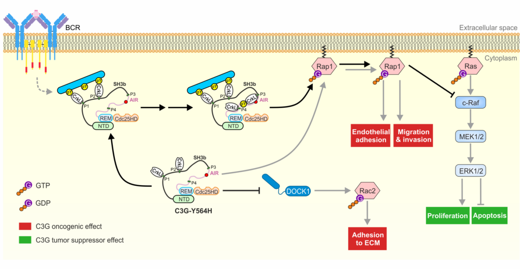
Schematic representation of the molecular pathways of the C3G protein.
Validation in animal models
Using animal models (mice), the human disease is simulated, and researchers study how cancer cells spread throughout the body under real physiological conditions. For this reason, the research team injected A20 cells into immunocompetent BALB/c mice. After 21 days, analyses revealed that the mice with C3G-Y564H cells developed more metastatic foci in the liver—smaller but more numerous—than the controls. This experiment confirmed the greater invasive capacity of the mutated C3G-Y564H cells. Clones with lower C3G expression showed even greater dissemination, validating the dual role of this protein.
Alba Morán-Vaquero (first author of the publication) notes: “These results in in vivo models validate the dual effect of C3G: it suppresses primary tumor growth via Rap1, but promotes metastasis by inhibiting Rac2 and reducing cell adhesion.”
Funding and Institutional Partnerships
The study is part of the Strategic Groups of the Regional Government of Castile and León and is funded by the Ministry of Science, Innovation, and Universities, ISCIII, the AECC Scientific Foundation, and ERDF funds. Researchers from the Salamanca Biomedical Research Institute (IBSAL), the Cancer Research Center (CSIC-USAL-FICUS), and Leiden University (Netherlands) are participating in the study.