Researchers from IBSAL, INCYL, and USAL are combining cell therapy and gene therapy to protect the brain against inflammation and DNA damage.
Bone marrow cells act as a “Trojan horse” to transport neuroprotective molecules to damaged areas of the nervous system.
The combination of cell therapy and gene therapy has made it possible to halt the loss of neurons in an experimental model of a neurodegenerative disease. The study, led by researchers from the Neuronal Plasticity and Neurorepair group at the Salamanca Biomedical Research Institute (IBSAL), the Castile and León Institute of Neurosciences (INCyL), and the University of Salamanca (USAL), shows that bone marrow cells genetically modified to produce higher levels of IGF-1— a protein with neuroprotective effects—are capable of reducing brain inflammation and protecting neurons from DNA damage.
The results, published in the high-impact international journal Journal of Tissue Engineering, open up new avenues for the development of advanced therapies for neurodegenerative diseases.
The study was conducted using a mouse model known as PCD (Purkinje Cell Degeneration), which replicates an inherited form of cerebellar ataxia. These animals suffer from rapid and aggressive degeneration of the cerebellar Purkinje cells—which are essential for controlling coordination and balance—and develop symptoms similar to those of human ataxias. In addition, they exhibit a progressive loss of neurons in other regions of the brain, such as the olfactory bulb, making this model a particularly valuable tool for studying various neurodegenerative processes within a single organism.
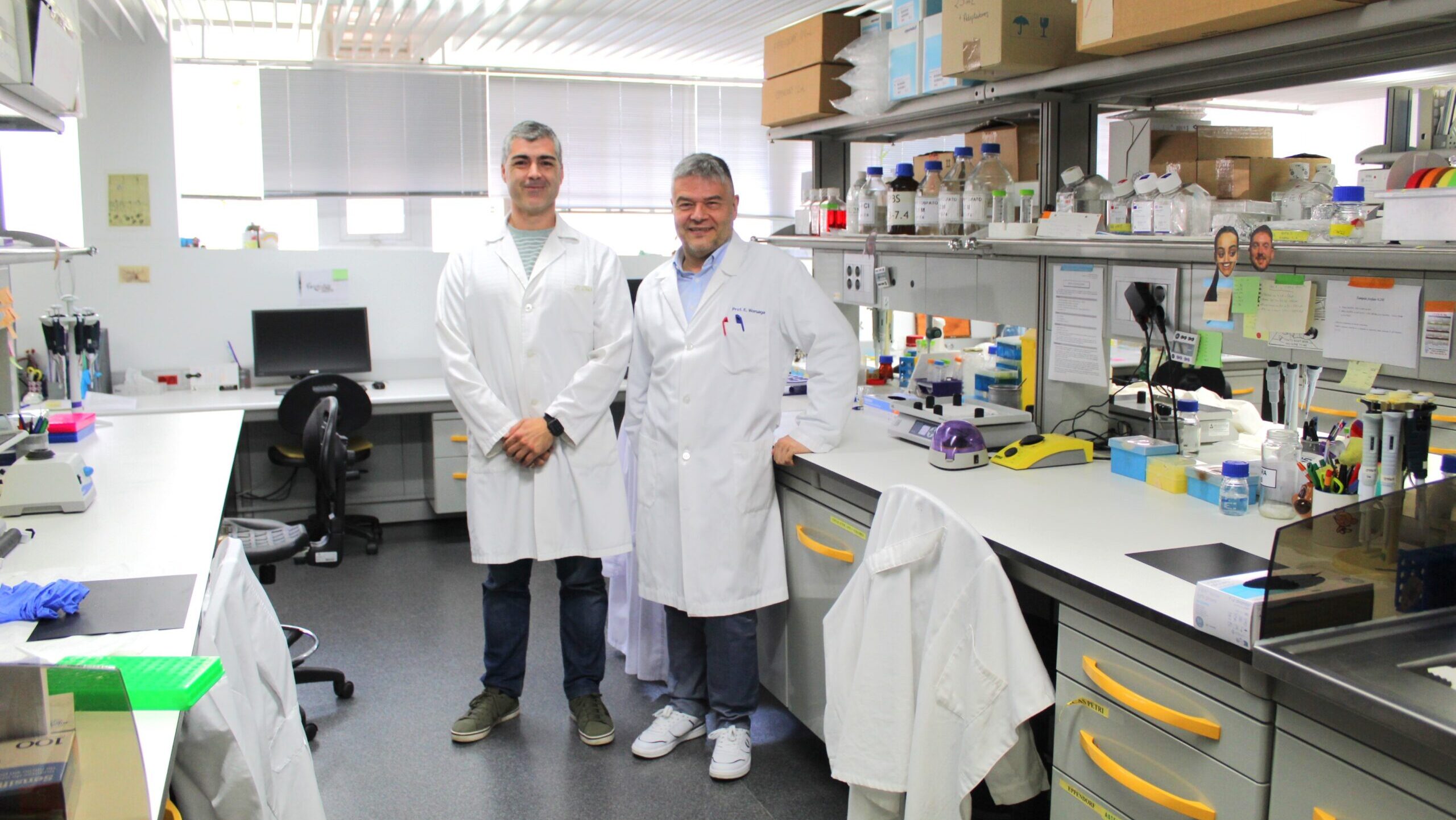
“The main advantage of this model lies precisely in the fact that it offers two different degenerative scenarios. While neuronal death in the cerebellum is extremely rapid, in the olfactory bulb it occurs more slowly, which provides a more favorable therapeutic window,” explains Eduardo Weruaga, professor of Cell Biology and Pathology at the University of Salamanca and principal investigator of the study.
A “Trojan horse” to deliver therapy to neurons
The group had already demonstrated in previous studies that bone marrow stem cell transplants were capable of reducing the neuroinflammation associated with neuronal degeneration. In this new study, they decided to take it a step further and turn those cells into therapeutic vehicles.
“We use bone marrow stem cells as a Trojan horse. We take advantage of their anti-inflammatory properties, but we also modify them so that they produce a molecule with neuroprotective properties, IGF-1. We wanted to achieve a dual effect,” says David Díaz, a full professor of Cell Biology at the University of Salamanca and co-director of the study.
IGF-1 (Insulin-like Growth Factor 1) is a growth factor naturally produced by the body and associated with the survival and maintenance of various tissues. Researchers had previously found that its levels were altered precisely in the brain regions affected by the disease. “What we do is enhance the body’s own molecules—molecules that play a role in the brain’s repair and maintenance processes,” adds Eduardo Weruaga.
To do this, they extracted hematopoietic cells from bone marrow and genetically modified them to produce large amounts of IGF-1. These cells were then transplanted into the animals.
The strategy revealed something unexpected. Although conventional transplants already reduced inflammation, the addition of IGF-1 provided an extraordinary additional effect. “What we saw was very striking. The death of these neurons was practically halted. The reduction was exceptional,” notes David Díaz.
The key lay in DNA repair
Initially, the researchers thought that the improvement might be due solely to a greater reduction in brain inflammation. However, they found that both conventional transplants and those enriched with IGF-1 had a similar anti-inflammatory effect. The explanation lay elsewhere.
“We saw that IGF-1 did something else. The neurons that were theoretically supposed to die showed hardly any DNA damage. That’s where the key really lay,” explains Díaz.
The study reveals that genetically modified cells activate cellular DNA repair mechanisms through a protein called IGFBP3, whose presence in the cell nucleus helps protect neurons from deterioration. This finding is one of the most novel aspects of the study and suggests that neuroprotection depends not only on controlling inflammation but also on preserving the genetic integrity of the cells.
The researchers also observed an unexpected finding in the cerebellum. Although degeneration in this region is much more aggressive and difficult to slow down, they found some surviving Purkinje cells in the treated animals—something they had virtually never seen in previous studies. “We were very surprised. Our target was the olfactory bulb, but when we analyzed the cerebellum, we found some Purkinje cells when normally none remain. There are only a few, but it’s a very striking finding,” says Eduardo Weruaga.
A line of research focused on slowing neurodegeneration
The Neuronal Plasticity and Neurorepair group , part of IBSAL and INCYL, has for years focused its research on understanding the mechanisms of neuronal death and developing strategies capable of slowing these processes.
“What we’re looking for are strategies that harness the body’s own molecules to boost the brain’s natural defense mechanisms,” explains Weruaga.
Although the results are very promising, the researchers emphasize that this is basic science conducted on animal models and that there is still a long way to go before any potential clinical application.
“Neuronal protection through cell therapy has a bright future, but it’s not something that will happen in the short term. The fact is that right now, for many neurodegenerative diseases, there are practically no treatment options,” says David Díaz.
This study is part of a broader line of research in which the group is seeking to identify other growth factors with neuroprotective properties. In fact, the researchers are already working with a new molecule, called VEGF-B, for which preliminary results are even more promising.
“The idea is to apply everything we’ve learned about IGF-1 and further enhance this strategy by using genetically modified cells,” David Díaz explains.
In addition, one of the upcoming challenges will be to analyze how these effects persist over the long term and to determine to what extent they may help protect the brain as we age.
Reference Article
Díaz D, Baranda-Alonso EM, Pérez-Revuelta L, Zapata-Acevedo JF, Torossian F, Anginot A, Alonso JR, Le Bousse-Kerdilès MC, Weruaga E. Genetically modified bone marrow cells halt mitral cell loss by modulating inflammation and protecting against DNA damage. Journal of Tissue Engineering. 2026;17:20417314261442787. DOI: 10.1177/20417314261442787.
IBSAL
The Salamanca Institute for Biomedical Research (IBSAL) was established on March 21, 2011, through an agreement signed by the Department of Health of the Regional Government of Castile and León and the University of Salamanca, which was joined in February 2012 by the Spanish National Research Council (CSIC). It is one of the 36 health research institutes accredited by the Carlos III Health Institute.
Its scientific activity is organized into six areas, comprising a total of 86 research groups: Cancer (22 groups); Cardiovascular, Renal, and Respiratory (11); Neuroscience (12); Infectious, Inflammatory, and Metabolic Diseases (19); Gene and Cell Therapy and Transplants (6); and Primary Care, Public Health, and Pharmacology (16).
For more information and contact details:
comunicacion@ibsal.es